Electron Microscopy
|
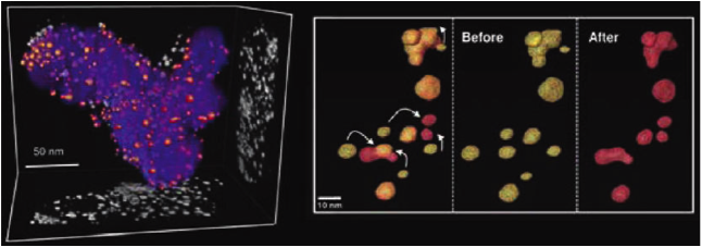
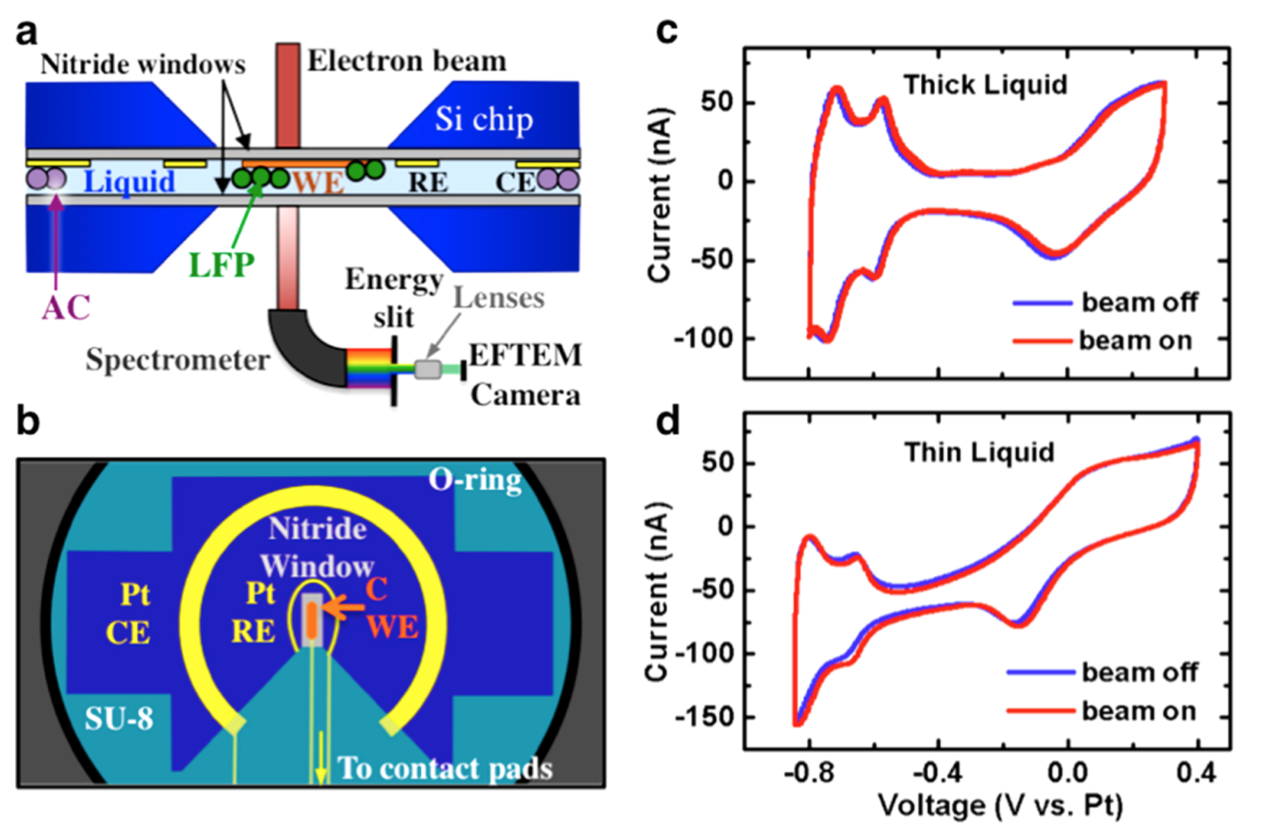
Electron microscopy provides tools to directly image the structure and chemistry of materials at atomic and nanometer length scales. The Abruña group uses advanced transmission electron microscopy (TEM) techniques to study materials for fuel cells and batteries. Identical-location imaging and tomography To understand the mechanisms of fuel cell catalyst degradation, the Abruña group uses identical-location TEM, where a TEM grid is loaded with catalyst particles and used as a working electrode for electrochemical aging, allowing the morphology of Pt-Co alloy and shape-controlled Pt catalysts to be observed before and after the experiment (Fig. 1). [1,2] Aberration-corrected Scanning TEM (STEM) imaging and electron energy-loss spectroscopy (EELS) Aberration-corrected STEM enables imaging of atomic structure at Angstrom resolution, and high beam current to enable rapid chemical imaging with EELS. The Abruña group uses these techniques to understand the detailed structure and performance of ordered intermetallic Pt3Co and dealloyed Cu3Pt fuel cell catalyst nanoparticles. [3,4] Operando electrochemical experiments in liquid The Abruña group has developed techniques to observe electrochemical processes in operation in the TEM using a specially designed, electron-transparent electrochemical cell (Fig. 2). This has allowed the observation of charging and discharging processes in LiFePO4, a lithium ion battery cathode material, using energy-filtered TEM to track the lithium ions (Fig. 3). [5,6] In-situ heating experiments The Abruña group has also performed in-situ heating TEM experiments to observe the dynamics of annealing and sintering in fuel cell catalyst nanoparticles. [7,8]
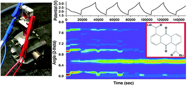
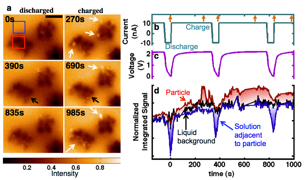
Figure 1. 3-D tomographic reconstruction of Pt-Co fuel cell catalyst before (yellow) and after (red) 30,000 electrochemical cycles between +0.6 V to +1.0 V (vs.RHE). Coalescence is revealed as the major degradation mechanism. Figure reproduced from [1]. Figure 2. Schematic of the in situ electrochemistry TEM holder and electrochemical data. (a) Cross-sectional view of the holder, with silicon nitride membranes encapsulating a fluid layer. The working electrode (WE), made of carbon, lies in the viewing window, with LiFePO4 (LFP) nanoparticles deposited on top. The platinum counter electrode (CE) is coated with an excess of activated carbon (AC). In EFTEM mode, energies are selected by a slit to be imaged. (b) Schematic of the top chip, with three patterned electrodes: a carbon WE on the viewing membrane, Pt reference electrode (RE), which is not used in the battery experiment, and Pt CE. The connection leads are covered by SU8, and the contact pads to the holder do not contact the liquid so as to minimize electrochemical activity outside the viewing window. The chips exhibited electrochemical activity qualitatively similar to that of an ex situ microelectrode, as shown for the Pt cyclic voltammetry (CV) in (c) and in (d). In extremely thin liquid layers (∼150 nm), the voltammetric profile exhibits a significant ohmic drop, as seen in (d). Reproduced from [6]. Figure 3. In situ charging and discharging of the cathode material LiFePO4 in 0.5 M Li2SO4 aqueous electrolyte. (a) The 5 eV spectroscopic EFTEM images of charging and discharging at indicated times. Scale bar is 400 nm. Bright regions are delithiated FePO4 and dark regions are LiFePO4. There are more bright regions of FePO4 at the end of charge cycles and less during the discharges. White arrows point toward “bright” charged particles, and black arrows point toward “dark” discharged particles. (b) Current profile corresponding to 10 C. Time = 0 s corresponds to the start of our study, which began on the third charge cycle (not on the first) after assembly. Arrows on the top axis indicate the times of the images shown in (a). The corresponding voltage profile is in (c), referencing the activated carbon counter electrode. (d) Integrated intensity over various regions, tracking with the voltage profile, from the regions shown by the boxes in (a). The solution becomes very dark during discharges and returns to the background level during charge. Regions of the particle are seen to light up and disappear, potentially due to delithiating and fracturing off of the particle cluster. During times when no imaging occurred, the data are linearly extrapolated, and for comparison, the intensity is brought to the same level by subtraction. Reproduced from [6]. Citations 1. Yu, Y., Xin, H. L., Hovden, R., Wang, D., Rus, E. D., Mundy, J. A., Muller, D. A. & Abruña, H. D. Three-dimensional tracking and visualization of hundreds of Pt−Co fuel cell nanocatalysts during electrochemical aging. Nano lett. 12, 4417-4423 (2012). |
Scanning Electrochemical Microscopy
|
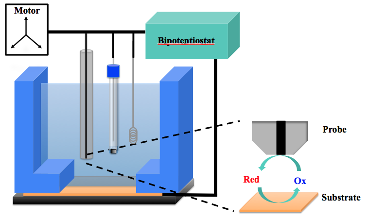
Scanning electrochemical microscopy (SECM) is a scanning probe technique in which an ultramicroelectrode (UME) is poised in close proximity to another electrode’s surface to obtain information about its reactivity—such as through feedback of a redox mediator in solution between the UME and surface (Figure 4)—or to modify the surface. The UME is attached to highly sensitive motors that can adjust the distance of the UME from the surface or move it laterally to obtain an image of the topography and/or reactivity of the surface. Our SECM system from CH Instruments (Figure 5) is used to probe the activity of electrode surfaces, such as graphene and photocatalysts.Our group has also combined SECM capabilities with in situ mass spectrometry for characterizing electrocatalysts.
Figure 4. Diagram of SECM cell and redox mediator feedback between the SECM tip and surface.
Figure 5. Picture of the Model 900 CH Instruments SECM in our group. Citations 1. N. L. Ritzert, J. Rodríguez-López, C. Tan, and H. D. Abruña. Kinetics of Interfacial Electron Transfer at Single Layer Graphene Electrodes in Aqueous and Non-Aqueous Solutions. Langmuir 2013, 29, 1683-1694. |

X-Rays
|
The Abruña group has over 25 years of experience in the in-situ and operando investigation of electrochemical interfaces using synchrotron radiation. These studies have been carried out locally at the Cornell High Energy Synchrotron Source (CHESS), as well as at the Advanced Photon Source (APS) at Argonne National Lab and the National Synchrotron Light Source (NSLS) at Brookhaven National Lab. Initial studies probed the underpotential deposition of metals and halogens on single-crystal precious-metal electrodes using X-ray standing waves. Recently, structural and chemical changes in several battery and fuel cell materials including metal oxides and nitrides, elemental sulfur, germanium nanowires, alkaline anion exchange membranes, and organic/organosulfur molecules were investigated using powder X-ray diffraction, X-ray absorption spectroscopy, small-angle X-ray scattering and inelastic X-ray scattering. By using multiple complementary techniques, deeper insight into the dynamics of the system can be gained. Future studies will introduce new techniques such as x-ray Raman scattering, X-ray emission spectroscopy and a variety of X-ray imaging techniques to the group’s x-ray toolbox. |